Midsagittal Section Of The Brain bmpwabbit

Brain, Midsagittal View Stock Image C024/9846 Science Photo Library
Midsagittal images of the brain provide a wealth of anatomic information and may show abnormalities that are pathognomonic for particular diagnoses. Using an anatomy-based approach, the authors identify pertinent anatomic structures to serve as a checklist when evaluating these structures. Subregions evaluated include the corpus callosum, pituitary gland and sellar region, pineal gland and.

Midsagittal Section Of The Brain bmpwabbit
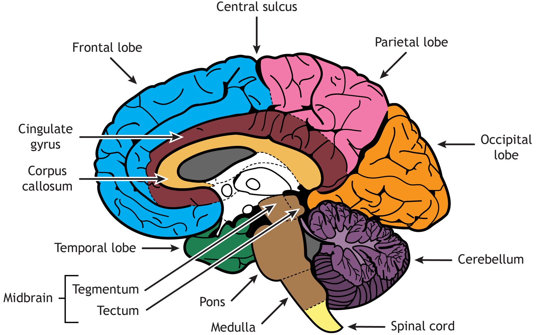
The midbrain, which can be seen only in this view, lies caudal to the thalamus, with the superior and inferior colliculi defining its dorsal surface or tectum (meaning "roof"); several midbrain nuclei, including the substantia nigra, lie in the ventral portion or tegmentum (meaning "covering") of the midbrain.

Sagittal View Of The Human Brain Labeled Sagittal Brain Transparent
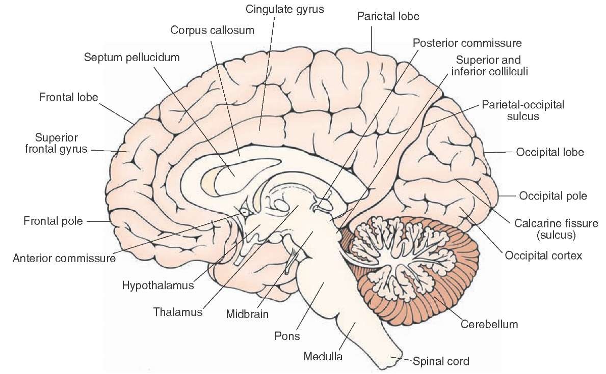
Figure 18.1. A midsagittal section of the brain. All four cerebral lobes are visible, as in the cingulate gyrus, which extends through the medial aspects of the frontal and parietal lobes. The corpus callosum sits beneath the cingulate gyrus. Below the cerebrum lies the midbrain, pons, medulla and cerebellum.

Midsagittal view of brain with labelled structures … Brain Anatomy
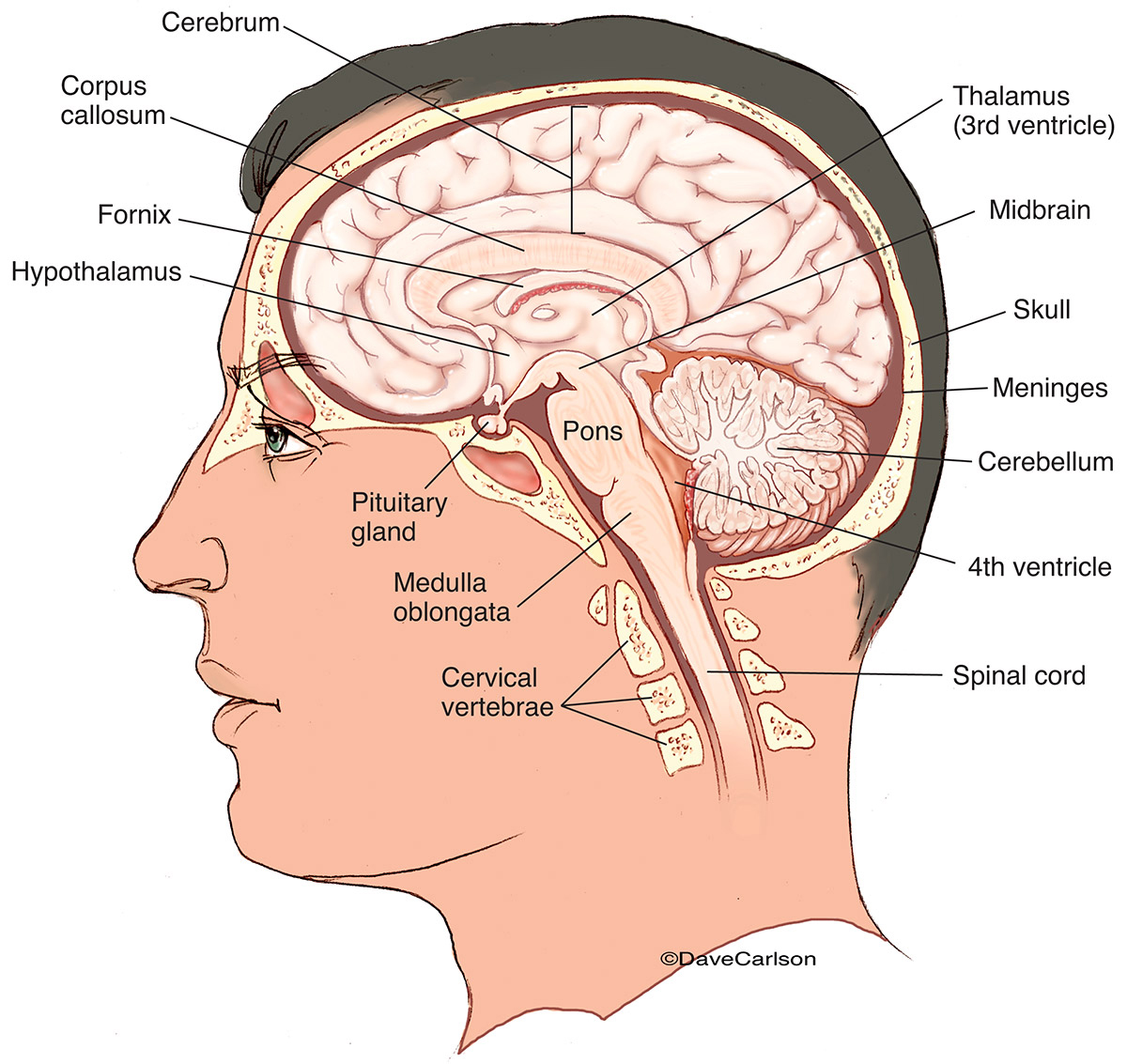
The brain is comprised of the cerebrum, cerebellum, and brainstem. The cerebrum is the most prominent region of the brain. It is divided into left and right hemispheres. The hemispheres have many of the same functions. For example, each perceives touch on one side of the body, but some functions demonstrate laterality, meaning they are.
Overview of the Central Nervous System (Gross Anatomy of the Brain) Part 1
Brainstem: Anatomy: The brainstem is divided into 3 sections: the midbrain (mesencephalon), the pons (metencephalon), and the medulla oblongata (myelencephalon) Function: The brainstem is responsible for swallowing, breathing, vasomotor control (blood pressure) the senses - taste, smell, hearing, touch, sight, and controlling heartbeat.
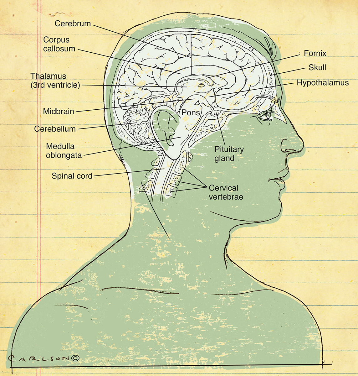
Human Brain Anatomy Midsagittal Carlson Stock Art
84 terms Preview Physiology Misc. Exam 1 Review 24 terms Preview Chapter 2 Anatomy and Physiology 89 terms js6jch8j62 Preview Terms in this set (75) Which organs are part of the central nervous system? brain and spinal cord
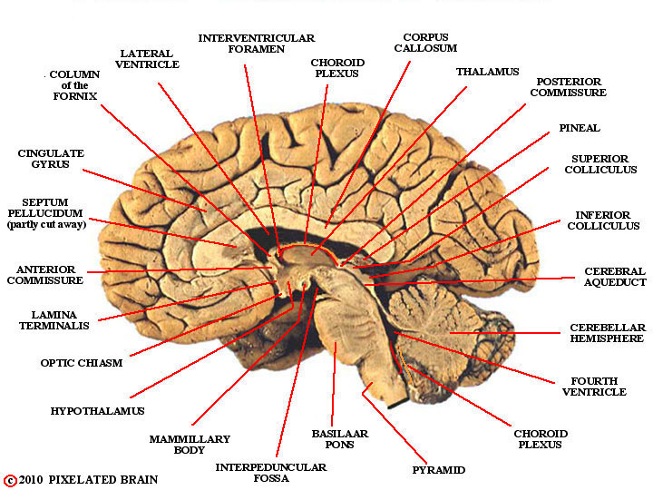
Pixelated Brain Module 14, Section 2 The four subdivisions of the
Torcula. Mid-sagittal view of the brain and ventricular system. The medial surface of the left cerebral hemisphere is in view. It is separated from the right hemisphere by the midline falx cerebri (largely removed) occupying the interhemispheric fissure. The lower free edge of this dural fold contains the inferior sagittal sinus, which enters.
Brain Structure Differentiation Introduction to Neuroscience
The anterior circulation. The four major arteries that arise from the internal carotid artery plus the posterior cerebral artery form the anterior circulation. The pattern of branching of each artery is similar: each gives rise to branches that supply cortical structures and each gives rise to branches that penetrate the ventral surface of the brain and supply deep structures (the basal.

Brain, Midsagittal View Stock Image C024/9844 Science Photo Library
Human Brain Anatomy MIDSAGITTAL VIEW OF BRAIN cerebrum Click the card to flip 👆 anterior part of the brain Click the card to flip 👆 1 / 14 Flashcards Learn Test Match Q-Chat Created by reneenicole13 Students also viewed midsagittal view brain 15.1 18 terms AnatomyPro123 Preview Exercise 20- Brain Structure and Function (D.) 5 terms Rachael27314
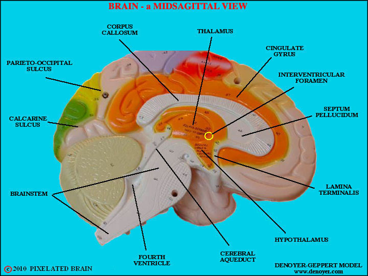
Pixelated Brain a model showing a midsagittal view of the brain
Anatomy | Major Parts of the Brain [Midsagittal View] Catalyst University 313K subscribers Subscribe 240 Share Save 16K views 3 years ago Anatomy | The Nervous System ⚡ Welcome to.

Midsagittal Section Of The Human Brain Chapter 12 The Central
LABEL THE BRAIN (MIDSAGITTAL VIEW) 3.0 (1 review) + − Flashcards Learn Test Match Created by sw33tpinkers Students also viewed Midsagittal View of The Brain 22 terms Anthony_Acevedo1 Preview Brain, midsagittal section labelling (car 19 terms Dajakewhite Preview Exercise 3 Oral Cavity, Throat, & Esophagus 5 terms leebrenda63 Preview

brain midsagittal view labels
the sagittal midline is observed when the cerebral aqueduct can be seen draining the third ventricle into the fourth ventricle. any displacement, of the cerebellar tonsils, or crowding of the foramen magnum. a review of the cisterns is important to note any displacement of the midline. moving superiorly, the cerebral aqueduct is observed for.

Midsagittal Section Of The Brain bmpwabbit
Vertebral Artery. Midsagittal section of the deep brain anatomy. This midline view demonstrates the third ventricle, with its roof formed by the body and column of the fornices and the velum interpositum. In the midline anteriorly, the lamina terminalis, optic chiasm and pituitary infundibulum are visible. The floor of the third ventricle is.
Midsagittal section of the brain Anatomy Kenhub
Lab 1 - Overview of the Human Brain Reading: Laboratory Guide, Lab 1 Coursera Media: Getting to Know the Human Brain Cerebral Cortex, Brainstem, and Blood Supply Basic Orientation in the Human CNS Finding the Central Sulcus Perspectives on neuroanatomy Neuroanatomy is a complex subject.

ANATOMY OF THE MEDIAL (MIDSAGITTAL) SURFACE OF THE BRAIN IN SITU
Series of annotated images of the sagittal midline of the brain in a normal participant. Image 1 : normal sagittal midline demonstrating the central venous vasculature and cisterns. 1. Great cerebral vein of Galen 2. Internal cerebral vein 3. Thalamostriate vein A. Cistern of the laminae terminalis B. Chiasmatic cistern C. Interpeduncular cistern
Human Brain Midsagittal View Carlson Stock Art
Step 1 Explanation: Labelled in order from top to bottom. 1. Corpus callosum 2. Choroid plexus of 3rd ventricle View the full answer Answer Unlock Previous question Next question Transcribed image text: Identify the structures labeled in this midsagittal view of the brain.
